
Retinal Tears / Vitreous Detachment
You or someone you know may have been diagnosed as having a retinal detachment. This disorder may lead to permanent loss of vision if not properly treated. We have prepared the following explanation to help you understand this condition better.
What is a retinal detachment?
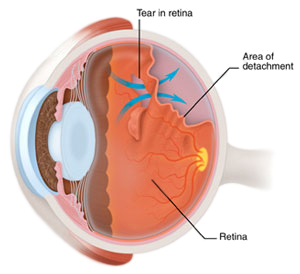
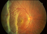 A retinal detachment occurs when the retina is pulled away from its normal position, lining the inside wall of the eye. The area of retina that is detached does not see light very well and the vision is blurred or lost. A retinal detachment may progress quickly and lead to complete loss of vision of the eye if not treated.
Treatment of retinal detachment
There are many ways to fix a retinal detachment. The decision of the type of retinal reattachment surgery and anesthesia depends upon the characteristics of your retinal detachment. Some retinal detachments require urgent or emergent surgery, while others may be postponed for a matter of days or weeks. Some retinal detachments do not require surgery.
- Scleral Buckle: Your doctor may recommend a scleral buckle operation. This may be performed exclusively or in conjunction with the vitrectomy mentioned below. In a scleral buckle operation your doctor will place a piece of silicone implanted material around all or part of your eye to indent the wall of the eye to counteract the force that led to the retinal tear and retinal detachment. Often your surgeon will drain the fluid underneath the detached retina to allow the retina to settle into its normal position more quickly. This operation is most commonly performed with local-sedation anesthesia (twilight) and may be performed either as an outpatient or inpatient operation.
- Vitrectomy: During a vitrectomy operation the vitreous jelly, which pulled on the retina to create the retinal tear, is removed from the eye and is usually replaced with a gas bubble. The gas bubble is eventually absorbed by the body and replaced with the eye's own internal fluids. A vitrectomy may be recommended when:
- diabetic retinopathy leads to the retinal detachment,
- hemorrhage or other opaque material prevents adequate view into the eye,
- the retinal tears are very far to the back of the eye,
- there is evidence of scar tissue on the surface of the retina, and
- in other conditions which may be associated with retinal detachment.